The cell culture environment is composed by the liquid in which cells are suspended (or surrounded by); the container where they are growing (well plate or similar, but also 3D structures like scaffolds), and the gas around them.
Cell culture media
Cells don’t only need medium in order to eat; they also interact with it to change according to the environmental conditions. The actual environment in the body provides a very rich and complex mixture of nutrients, proteins and oxygen that is impossible to replicate artificially. However, there are ways to provide cells with the required compounds for them to maintain, duplicate or differentiate. Maintenance requires the presence of glucose or other primary sources of energy, oxygen and sometimes a matrix on which to grow. Growth, unless you’re dealing with highly proliferative cells, generally requires the addition of growth factors. Growth factors are proteins that are naturally present in the body and promote cellular duplication. Differentiation can happen spontaneously in some cases, but in general differentiation cues are provided in the form of chemicals or proteins to help mature cells towards the desired lineage.
Types of cell culture media
There are three broad types of medium sources: serum-free medium, serum-containing medium and defined medium. Serum is a protein-rich liquid that is extracted generally from cows (you don’t want to know how) and that is very good at enhancing growth, because most of the required proteins are there. It is expensive and should be handled with care (a mere drop on your skin can give you an itchy feeling for a bit). Conversely, serum-free medium is formulated to contain all the required proteins, but they have been added in specific amounts (its composition is known). It can still contain elements extracted from animal sources, unlike defined medium which only contains plant and microorganism-derived proteins. Research tends to move towards using serum-free and defined medium as it helps for regulation purposes if planning to use cells medically (this is known as GMP compliance). Of course, serum-containing medium doesn’t contain only serum. It is generally mixed with vitamins, glucose, glutamate and other components that promote growth, which are all contained in standard synthetic formulated medium such as IMDM or RPMI.
Incubator conditions
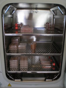
Incubators have two main purposes: keeping the temperature constant at 37ºC (or the one required by your cells) and keeping the oxygen and carbon dioxide concentrations at the correct levels (20% O2 and 5% CO2 is the most common). For some experiments, hypoxic conditions (with oxygen levels much lower than the atmospheric 20%) are used, because they are believed to be closer to physiological conditions. Many experiments under hypoxia are performed at 5% or lower oxygen concentrations. To achieve this, incubators have a heating system and a CO2/N2 pumping system, connected to temperature and CO2/O2 sensors. CO2 is delivered from gas cylinders and oxygen concentrations can be adjusted using a nitrogen cylinder (since 20% or lower O2 concentrations are always used, the aim is to reduce the ambient air concentration of oxygen).
Humidity is maintained by using a water tray which needs to be monitored and changed every 1-2 weeks. When changing this water, ensure you use purified water and you add disinfectants (Aquaguard) so microorganisms do not proliferate there. Generally, cells of the same origin can be kept in the same incubator, preferably in different shelves/trays. Items should be placed with extreme care not to move other items that are already in, as for example well plate lids can remain slightly open and spinner flasks might fall. Always remember to close the inner door properly before closing the outer door. Never take items out that do not belong to you, even if just to make space. If there is a spillage, clean it up asap with Virkon and then ethanol. Be quick placing items in or taking items out, as the environmental conditions inside the incubator change quickly due to the entrance of air at a different temperature and with a different composition.
Monitoring incubator conditions
Incubator conditions are regularly checked by using secondary measurements to verify the primary sensor is accurately reporting temperature or gas concentration. A simple thermometer dipped in a sterile tube with purified water (and some antibiotics) can be permanently left in the incubator and checked at least once a week. It should be kept in an area where it doesn’t obstruct the manipulation of flasks, ideally towards the back. CO2 concentration is measured with a special analyzer (FYRITE or an electronic device) also on a weekly basis. It is common practice to note down the weekly measurements on a paper placed on the incubator door so all users know it is working under normal conditions. CO2 cylinder pressure should be checked regularly as well so it doesn’t run out when no one is is the lab to change cylinders. If using nitrogen cylinders to adjust O2 concentration, their pressure should be checked as well quite often (twice a week, ideally once a day) as they run out quite quickly.
Cleaning the incubator
Incubators should be cleaned a minimum of once a month. Any cultures running inside should be placed in another incubator temporarily until the cleaning is over. Some incubators are self-cleaning, so you only need to set it up when it’s more convenient. Traditional incubators require autoclaving of the removable parts and thorough cleaning of the inside walls with Virkon and ethanol. Once the removable parts are put back, the whole should be UV’ed overnight by leaving the UV light on inside it and a clear sign on the door, so no one opens it. The incubator is then ready to be used again, and the cultures can be placed back.
If you’re using any machines inside the incubator that need cables to be plugged outside (such as a shaker), make sure they do not prevent the door from being closed.
Cell culture flasks and well plates
T-flasks
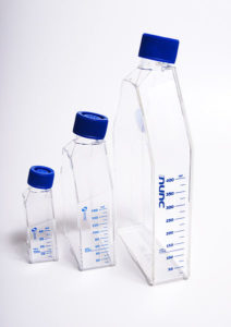
Cells are maintained in disposable flasks or reusable reactors (usually made out of glass). Disposable flasks come essentially in two different types: the ones for static culture (T-flasks and well plates) and the ones for shaked culture (erlenmeyers and spinner flasks). T-flasks are used when expanding cells for later testing or when larger amounts of cells are required in a single test. They come in several standard sizes: T-25, T-75 and T-175.
| Flask | Surface | Volume range |
|---|---|---|
| T-25 | 25 cm2 | 5-10 mL |
| T-75 | 175 cm2 | 15-25 mL |
| T-175 | 175 cm2 | 40-70 mL |
Generally, T-25 flasks are used when starting a new culture from frozen cells, which are then passaged and transferred into a T-75 flask to increase the volume of medium due to the increase in cell number, and finally T-175 flasks are useful for those experiments where you need large amounts of cells.
Well plates
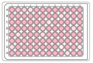
Well plates come in multiples of 6 wells: 6-, 12-, 24-, 48-, 96-, 384-well plates… They must have a lid so they can be placed in an incubator and avoid any contamination, but some of them (especially 96-well plates) are commonly used for analyses and are sold without one. Below is a reference table for well plate sizes and volumes:
| Number of wells per plate | Surface | Volume range |
|---|---|---|
| 6 | 9.5 cm2 | 2-5 mL |
| 12 | 3.8 cm2 | 0.8-1.5 mL |
| 24 | 1.9 cm2 | 0.4-1 mL |
| 48 | 0.45 cm2 | 0.2-0.5 mL |
| 96 | 0.32 cm2 | 0.36 mL |
| 384 | 0.056 cm2 | 0.15 mL |
Both wells and flasks can have their bottom inner surface coated to enhance cell adhesion in adherent cell lines. Sometimes specific cell lines require a particular coating in which case you will have to purchase special flasks for your own experiments, check in the cell line protocol.